Compact Bone Diagram Microscope : Chapter 6 Page 7 Histologyolm / The cells of compact bone, which is also called cortical bone, appear to be tightly packed into a solid mass.
Compact Bone Diagram Microscope : Chapter 6 Page 7 Histologyolm / The cells of compact bone, which is also called cortical bone, appear to be tightly packed into a solid mass.. However, compact bones also serve a function in storing and releasing calcium to the. The central canal, lamellae, canaliculi, and lacunae with osteocytes are apparent. Under magnification you can clearly see the system of concentric circles that forms compact bone. It is thick and dense. This human bone section shows the haversian canal (or osteon) structure of compact bone tissue.
Each group of concentric circles (each tree) makes up the microscopic structural unit of compact bone called an osteon (this is also called a haversian system). The scanning electron microscope (sem) is among the most frequently used instruments for examining bone. The darker ring consists of layers of bone matrix made by cells called. Compact bone is formed in concentric circles. The compact bone is the main structure in the body for support, protection, and movement.
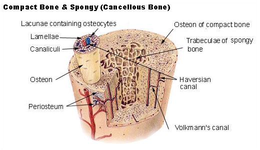
Compact Bone Spongy Bone And Other Bone Components Human Anatomy And Physiology Lab Bsb 141 from cnx.org Spongy bone sharpey's fibers compact bone blood vessel periosteum perforating (volkmann's) canal blood vessel figure 7.3 microscopic structure of compact bone. Compact bone histology slide structure with diagram. Under the microscope, bone can be divided into two types compact bone forms the outer 'shell' of bone. This type of bone is located between layers of compact bone and is thin and porous. It is enveloped by lamellae in the ground substance, which may be more or less impregnated with silver nitrate. Do you want to learn the details of the histology of compact bone with labelled diagram and authentic slide images? Under the microscope, bone can be divided into two types compact bone forms the outer 'shell' of bone. Bone, dried ground preparation, human.
It is enveloped by lamellae in the ground substance, which may be more or less impregnated with silver nitrate.
Compact bone histology slide structure with diagram. Bone tissue and cells under the microscope introduction. 0 0000 a shoutout is a way of letting people know of a. Compact and spongy bone with dr. There are small canals that run through the bone, which allow blood vessels to penetrate it. It is lined by the endosteum and is filled with yellow bone marrow. Before placing your slide on the microscope stage, remember to read the label, examine the slide with your eye and note any visible macroscopic features that might help your examination. Transverse section of an osteon with its haversian canal [1. Each group of concentric circles (each tree) makes up the microscopic structural unit of compact bone called an osteon (this is also called a haversian system). The compact bone is composed of calcified extracellular material the bone matrix and 3 major cell types which are osteoblast which ssynthesize and secrete the organic components of bone matrix which include type 1 collagen fibers proteoglycans and several glycoproteins such as ostepnectin. Bone, dried ground preparation, human. You can think of compact bone as being very similar. Learn vocabulary, terms, and more with flashcards, games, and other study tools.
The darker ring consists of layers of bone matrix made by cells called. This is the area of bone to which ligaments and tendons attach. In the lightly stained areas, their orientation. Start studying compact bone microscopic labeling. Spongy bone sharpey's fibers compact bone blood vessel periosteum perforating (volkmann's) canal blood vessel figure 7.3 microscopic structure of compact bone.
A C Ultrastructure Of Bone A Light Microscope Image Of A Ground Download Scientific Diagram from www.researchgate.net The darker ring consists of layers of bone matrix made by cells called. A diagram of the anatomy of a bone, showing the compact bone. The compact bone is the main structure in the body for support, protection, and movement. Compact bone, also called cortical bone, dense bone in which the bony matrix is solidly filled with organic ground substance and inorganic salts, leaving only tiny spaces (lacunae) that contain the osteocytes, or bone cells.compact bone makes up 80 percent of the human skeleton; The compact bone is composed of calcified extracellular material the bone matrix and 3 major cell types which are osteoblast which ssynthesize and secrete the organic components of bone matrix which include type 1 collagen fibers proteoglycans and several glycoproteins such as ostepnectin. Under magnification you can clearly see the system of concentric circles that forms compact bone. Bone, dried ground preparation, human. Like other tissues in the body, bones are made up of specialized cells that serve different functions.
Start studying compact bone under microscope.
However, compact bones also serve a function in storing and releasing calcium to the. Trabecular bone, also known as cancellous bone or spongy bone, mainly serves a metabolic function. These bones tend to support weight and. You can think of compact bone as being very similar. Although the calls are close together, this type of bone is not completely solid. Spongy bone is used for more active functions of the bones, including blood cell production and ion exchange. The darker ring consists of layers of bone matrix made by cells called. Compact bone, or cortical bone, mainly serves a mechanical function. Lamellar bone makes up the compact or cortical bone in the skeleton, such as the long bones of the legs and arms. Under magnification you can clearly see the system of concentric circles that forms compact bone. Compact bone is formed in concentric circles. Learn vocabulary, terms, and more with flashcards, games, and other study tools. Some, mostly older, compact bone is remodelled to form these haversian systems (or osteons).
Online quiz to learn compact bone microscope slide labeled ; Compact and spongy bone with dr. Lamellar bone makes up the compact or cortical bone in the skeleton, such as the long bones of the legs and arms. Each group of concentric circles (each tree) makes up the microscopic structural unit of compact bone called an osteon (this is also called a haversian system). Some, mostly older, compact bone is remodelled to form these haversian systems (or osteons).
Seer Training Structure Of Bone Tissue from training.seer.cancer.gov Learn vocabulary, terms, and more with flashcards, games, and other study tools. The diagram above shows a longitudinal view of an osteon. A photo taken through a microscope that shows the anatomy of compact bone with a detailed view of an osteon. Start studying compact bone under microscope. At this level of magnification, the fundamental structure of compact bone is visible. However, compact bones also serve a function in storing and releasing calcium to the. Compact bone, or cortical bone, mainly serves a mechanical function. Compact bone is very strong, and it provides the supportive strength of long bones.
Transverse section of an osteon with its haversian canal [1.
Compact bone histology slide structure with diagram. (b) in this micrograph of the osteon, you can clearly see the concentric lamellae and central canals. A diagram of the anatomy of a bone, showing the compact bone. Under the microscope, bone can be divided into two types compact bone forms the outer 'shell' of bone. (the inset shows a more highly magnified view.) notice the position of osteocytes in lacunae (cavities in the matrix). The darker ring consists of layers of bone matrix made by cells called. At this level of magnification, the fundamental structure of compact bone is visible. Like other tissues in the body, bones are made up of specialized cells that serve different functions. Each group of concentric circles (each tree) makes up the microscopic structural unit of compact bone called an osteon (this is also called a haversian system). Start studying compact bone under microscope. Due to the strong nature of compact bone, compared to spongy bone, it is the preferred tissue for strength. The collagen fibers in the more heavily stained lamellae are arranged in a circular fashion; To prepare this slide, a bone specimen is ground thin and then.


0 Komentar